| |
|
|
Multi-Speciality Focus
We cover all medical needs, from hair transplants to heart transplants
-
-
-
-
-
-
-
-
-
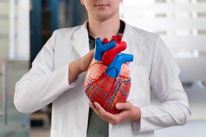
Cardiology
Cardiology plays a pivotal role in maintaining heart health, covering everything from regular check-ups to complex procedures. We partner with esteemed cardiologists from top hospitals. Our team employs cutting-edge technology and is committed to protecting your heart, the most essential organ. Whether detecting a faint murmur or performing sophisticated bypass surgery, our specialists approach every situation with meticulous attention and heartfelt concern, ensuring your heart stays robust and steady.
Cardiac Treatments
Our Cardiological Services
Ablation therapy is a minimally invasive procedure employed by healthcare professionals to remove abnormal tissue associated with various conditions. This technique can be used to address irregular heart rhythms as well as tumors in areas such as the lungs, breast, thyroid, liver, and other parts of the body.
Types of Ablation Therapy
Ablation of Atrial Fibrillation: This procedure addresses irregular heartbeats by creating tiny scars in the heart tissue to help restore normal rhythm.
Cardiac Ablation: Utilizing heat or cold energy, this method creates small scars within the heart, aiding in the restoration of a regular heartbeat.
Cryoablation for Cancer: This technique involves freezing cancerous tissue with a cryoprobe that is inserted directly into the tumor.
Laser PVP Surgery: A minimally invasive approach for treating an enlarged prostate, this procedure uses laser technology to vaporize excess prostate tissue.
Radiofrequency Neurotomy: This treatment temporarily disrupts specific nerves' ability to transmit pain signals by applying heat generated from radio waves.
Your heart contains four valves: the mitral, tricuspid, aortic, and pulmonic valves. The aortic valve is situated between the left ventricle (the heart's lower pumping chamber) and the aorta, the largest artery in the body. These valves ensure that blood flows in a single direction through the heart.
Aortic valve surgery
There are two primary surgical options for the aortic valve: Repair and Replacement. During the surgery, which may also involve the aorta, the aortic valve can either be repaired or replaced. The decision on whether to repair or replace the valve is based on diagnostic test results, heart structure, age, existing health conditions, and other factors. Aortic valve surgery can be performed using traditional methods or through minimally invasive techniques.
Symptoms of aortic valve disease.
Many individuals with aortic valve disease may not show any symptoms, even in cases of severe stenosis (narrowing) or insufficiency (leakage). Early signs of aortic valve disease typically include:
- Fatigue
- Easy exhaustion
- Decreased energy
- Swelling in the ankles
- Palpitations (extra or missed heartbeats)
As the condition progresses, more serious symptoms may develop, such as:
- Shortness of breath
- Chest pain
- Dizziness or fainting
Coronary Artery Bypass Grafting (CABG), commonly known as heart bypass surgery, is a procedure that creates alternative pathways for blood to flow to the heart by bypassing blocked coronary arteries using grafts from the patient's own arteries and veins. Traditionally, this surgery is performed on a still heart, with the heart-lung machine managing circulation during the operation. However, advancements in medical technology have led to a groundbreaking technique called Off-Pump Surgery. In Off-Pump CABG, the surgery is conducted while the heart remains beating, utilizing specialized equipment to maintain a controlled environment around the heart. This method aims to lower the risk of complications such as renal failure and stroke, as well as reduce the need for blood transfusions. The choice between on-pump and off-pump CABG is determined by the surgeon after a thorough assessment of the patient's circulatory system, typically made during the surgery itself based on the individual's medical condition.
Types of CABG
There are two primary types of grafts used in CABG: Arterial and Vein Grafts.
Arterial Grafts
Arterial grafts can be divided into two main types:
- Internal Thoracic Artery: This is a commonly used graft, also known as the internal mammary artery (IMA). There are two internal thoracic arteries in the chest that are preserved at their origin due to their supply of oxygen-rich blood. One end is cut and connected to the coronary artery below the blockage.
- Radial Artery: This is another frequently used arterial graft. Before utilizing a radial artery, various tests are conducted to ensure it's the best option. After surgery, patients may be prescribed calcium channel blockers for several months to help keep the artery open. It's common to experience numbness in the wrist following this procedure.
Vein Graft
The saphenous veins in the legs can be used as bypass grafts through a technique called minimally invasive saphenous vein removal. This method involves making one or two small incisions at the knee and a smaller incision at the groin, resulting in quicker recovery and less scarring compared to traditional surgical methods. However, these vein grafts tend to have a shorter lifespan compared to arterial grafts.
Coronary angioplasty, also referred to as percutaneous coronary intervention (PCI), is a minimally invasive procedure aimed at opening narrowed or blocked coronary arteries, thereby restoring blood flow to the heart muscle. Our advanced facility provides comprehensive coronary angioplasty services, delivered by a team of skilled cardiologists and cardiac specialists.
Understanding Coronary Angioplasty
Coronary angioplasty is an effective treatment for coronary artery disease, a condition characterized by plaque buildup in the coronary arteries. This procedure helps alleviate symptoms such as chest pain (angina) and shortness of breath by enhancing blood flow to the heart.
Key Steps in the Coronary Angioplasty Procedure
The coronary angioplasty procedure typically involves the following steps:Preparation: The patient is prepared in a dedicated catheterization laboratory (Cath Lab), where local anesthesia is administered at the incision site, usually in the leg, arm, or wrist.Catheter Insertion: A catheter is introduced into a blood vessel and guided toward the blocked coronary artery, using live X-ray imaging (fluoroscopy) for assistance.Blockage Identification: Contrast dye is injected through the catheter to help visualize the blockage in the coronary artery.Balloon Inflation: A balloon-tipped catheter is maneuvered to the blockage site and inflated to compress the plaque and expand the artery.Stent Placement: In many instances, a stent-a small wire-mesh tube-is placed in the artery to offer structural support and keep it open. Drug-eluting stents (DES) are coated with medication to help prevent the artery from re-narrowing.Completion: After successfully treating the blockage and placing the stent, the balloon is deflated, and the catheter is withdrawn.
Stents are frequently used alongside coronary angioplasty to maintain openness in narrowed coronary arteries. A stent is a small, wire-mesh tube that acts as a scaffold, preventing the artery from collapsing after it has been widened. Some stents, known as drug-eluting stents (DES), are coated with medication that gradually releases to help prevent the artery from re-narrowing. In contrast, stents without a medication coating are referred to as bare metal stents (BMS).
Why Is a Stent Necessary
Patients may require a stent during emergency procedures involving blocked coronary arteries, allowing surgeons to perform angioplasty to restore blood flow. Stents can also be employed in other situations, such as preventing aneurysms in the brain, aorta, or other blood vessels from rupturing. They can be used to open various passageways, including:
- Bile Ducts: These tubes transport bile to and from the digestive organs, facilitating digestion.
- Bronchi: The small airways within the lungs that help direct air to the alveoli for gas exchange.
- Ureters: The tubes that connect the kidneys to the bladder, allowing urine to flow from the kidneys.
Stenting can significantly improve quality of life by alleviating symptoms such as chest pain (angina) and shortness of breath. The procedure is minimally invasive, often leading to shorter recovery times and less hospital stay compared to traditional surgery. Regular follow-up care is essential to monitor the function of the stent and ensure optimal heart health.
A pacemaker is a compact electronic device implanted in your chest, just below the collarbone, designed to help regulate your heart rate. It is typically recommended for individuals experiencing dangerously low heart rates, a condition known as bradycardia. By sending electrical impulses to the heart, a pacemaker helps ensure it beats at a normal rate, improving blood flow and overall heart function.
Types of pacemakers
There are three main types of pacemakers:
- Single Chamber Pacemakers: These consist of a single lead connected to one heart chamber, usually the right ventricle. This type is often used when the heart's upper chambers are functioning properly.
- Dual-Chamber Pacemakers: These have two leads that connect to both the atrium (upper chamber) and the ventricle (lower chamber). This configuration allows for better coordination of heart contractions, which can be beneficial for patients with specific rhythm issues.
- Biventricular Pacemakers: Also known as cardiac resynchronization therapy (CRT) devices, these pacemakers have three leads that stimulate both ventricles simultaneously. This approach is particularly useful for patients with heart failure, as it helps improve the efficiency of the heart's pumping action.
The LVAD is a battery-operated mechanical pump that is surgically implanted to assist a heart that cannot effectively pump blood on its own. This device typically includes a tube that draws blood from the left ventricle into the pump, which then sends it into the aorta, the main blood vessel exiting the left ventricle. This process helps support the weakened ventricle. The pump is positioned in the upper abdomen, with another tube extending through the abdominal wall to connect to an external battery and control system.
Major Indications:
- Initially designed as a "bridge to transplant," LVADs are now also used for long-term therapy.
- Indicated for patients with Stage D heart failure and Class IV heart failure.
- Increasingly utilized as destination therapy for patients with end-stage heart failure when heart transplantation is not a viable option.
Transcatheter Aortic Valve Replacement (TAVR) is an innovative technique for treating aortic valve disease. This minimally invasive option is designed for patients at moderate, high, or extreme risk for traditional open-heart surgery. During the procedure, the diseased aortic valve, which does not open properly, is replaced with a new valve, restoring blood flow and alleviating the symptoms associated with aortic stenosis.
Benefits of TAVR
- It enhances the quality of life for patients with aortic stenosis who are unable to undergo surgery or where surgery poses significant risks.
- TAVR can lower the risk of mortality.
- The procedure alleviates symptoms of aortic valve stenosis and contributes to improved overall health.
- Patients typically enjoy faster recovery times and can often return to their normal activities more quickly.
Neurosurgery
Neurosurgery is dedicated to addressing conditions related to the brain and nervous system. We collaborate closely with skilled neurosurgeons from our partner hospitals who specialize in the intricate field of brain surgery and comprehensive neurological care. Utilizing state-of-the-art technology and a profound knowledge of the brain, they are equipped to diagnose and treat a variety of neurological disorders, ensuring that your cognitive health and overall well-being are in capable hands.
Treatment for Arteriovenous Malformations (AVMs) generally involves surgical removal, embolization (blocking the abnormal blood vessels), or stereotactic radiosurgery (focused radiation therapy) to lower the risk of rupture and address symptoms such as seizures or neurological deficits. The choice of treatment depends on factors like the AVM's location, size, and associated symptoms.
Steps in Treating Arteriovenous Malformations (AVMs)
- Diagnosis and Assessment: AVMs are identified using imaging techniques such as MRI, CT angiography, or cerebral angiography to evaluate their location, size, and blood flow patterns.
- Treatment Planning: Based on the AVM's characteristics (including size, location, and symptoms), treatment options are discussed. This planning often involves a multidisciplinary team that may include neurosurgeons, interventional radiologists, and neurologists.
- Intervention: Treatment methods may include:
- Surgical Resection: Removal of the AVM.
- Endovascular Embolization: Injecting a substance to block the abnormal blood vessels.
- Stereotactic Radiosurgery: Using focused radiation to shrink or close off the AVM.
- Follow-Up and Monitoring: Post-treatment, patients are closely monitored to evaluate the effectiveness of the intervention and to check for any complications or recurrence. Follow-up imaging and clinical assessments are typically scheduled to ensure that the AVM remains controlled.
Neurosurgery involves complex procedures related to the brain, spinal cord, skull, and spinal column, requiring specialized expertise. With advancements in technology transforming healthcare, significant strides have been made in neuro care, leading to improved diagnostic techniques and treatment outcomes.
What Are Tumors?
Tumors are abnormal tissue growths that can form in various parts of the body, including the brain. These growths can be classified as benign (non-cancerous) or malignant (cancerous). Benign tumors generally grow slowly and may not invade surrounding tissues, while malignant tumors can grow rapidly and may spread to other parts of the body.
Types of Brain Tumors:
Brain tumors can be classified into primary tumors, which originate in the brain, and secondary tumors, which spread from other parts of the body. Common types include gliomas, meningiomas, and pituitary adenomas.
Symptoms:
The symptoms of brain tumors can vary widely depending on their location, size, and growth rate. Common symptoms may include headaches, seizures, vision changes, cognitive difficulties, and motor function impairments.
Treatment Options
- Surgery: Surgical removal of the tumor is typically the primary treatment for brain tumors, with the goal of resecting as much of the tumor as possible while preserving neurological function.
Radiation Therapy: Radiation therapy employs high-energy beams to target and destroy cancer cells. It can be used as a standalone treatment or in conjunction with surgery and/or chemotherapy.
Chemotherapy: Chemotherapy involves the administration of drugs that kill cancer cells or inhibit their growth. This treatment can be delivered either orally or intravenously, depending on the specific medication and patient needs.
Targeted Therapy: Targeted therapy uses drugs designed to specifically attack cancer cells while minimizing harm to healthy cells. These therapies often interfere with specific molecules involved in tumor growth and progression, providing a more focused approach to treatment.
Advancements in imaging technology, such as MRI and CT scans, have improved the ability to diagnose and monitor brain tumors, allowing for more personalized and effective treatment plans.
A craniotomy is a surgical procedure that involves removing a portion of the skull to expose the brain. Surgeons utilize specialized tools to extract a segment of bone known as a “bone flap,†which is temporarily removed and later replaced once the brain surgery is finished.
Craniotomy procedures typically consist of the following steps:
- Incision: A scalp incision is made to access the skull bone.
- Bone Flap Removal: A section of the skull bone (bone flap) is carefully extracted using specialized instruments to gain access to the brain.
- Brain Exposure and Surgery: The neurosurgeon opens the protective layer covering the brain (the dura) and performs the required surgical procedure, which may involve tumor removal, vascular repairs, or other interventions.
- Closure: After the surgery is completed, the bone flap is repositioned and secured with plates, and the scalp incision is closed with sutures or staples.
These steps are designed to provide safe access to the brain while minimizing tissue damage and facilitating recovery.
Deep Brain Stimulation (DBS) is a specialized neurosurgical procedure designed to alleviate symptoms of movement disorders, including Parkinson's disease, essential tremor, dystonia, epilepsy, and obsessive-compulsive disorder. During the procedure, small electrodes are implanted in targeted areas of the brain to deliver electrical stimulation. This stimulation helps regulate abnormal brain activity and reduces symptoms that do not respond to medication or other treatments.
Endoscopic skull base tumor surgery involves using a small tube equipped with a camera, inserted either through the nose or a small incision in the skull, to excise tumors located at the base of the skull, such as pituitary tumors. This approach causes less tissue disruption and allows for faster recovery compared to traditional surgical methods. The typical procedure for endoscopic skull base tumor surgery includes the following steps:
Preparation: The patient is positioned and prepped for surgery, which may involve general anesthesia. The surgeon may utilize imaging techniques, such as MRI or CT scans, to accurately pinpoint the tumor's location.
Endoscopic Approach: A flexible endoscope with a camera and light source is introduced through the nostril or a small incision in the skull, providing a clear view of the tumor and surrounding structures.
Tumor Removal: Utilizing specialized surgical instruments inserted through the endoscope, the surgeon meticulously excises the tumor, ensuring minimal damage to adjacent healthy tissues, including nerves and blood vessels.
Closure and Recovery: After the tumor is successfully removed, any incisions are closed, usually with dissolvable stitches. Patients are closely monitored during the post-operative period to ensure proper healing and to address any discomfort or complications.
Epilepsy surgery is a surgical intervention designed to control or significantly reduce the frequency of seizures in individuals with epilepsy.
Types of Epilepsy Surgery
- Anatomical or Functional Hemispherectomy: A procedure that involves the removal or disconnection of one hemisphere of the brain to control seizures.
- Corpus Callosotomy: Surgery that involves cutting the corpus callosum, which connects the two hemispheres of the brain, to prevent the spread of seizures.
- Focal Resection: Removal of a specific area of the brain where seizures originate, aimed at reducing or eliminating seizure activity.
- Laser Interstitial Thermal Therapy (LITT): A minimally invasive technique that uses laser energy to destroy brain tissue responsible for seizures.
- Lesionectomy: Surgical removal of a brain lesion or abnormality that contributes to seizure activity.
- Multiple Subpial Transections: A technique that involves making cuts in the brain's surface to interrupt seizure pathways without removing tissue.
- Neurostimulation Device Implantations: Implantation of devices, such as a vagus nerve stimulator, to help regulate brain activity and reduce seizures.
- Stereotactic Radiosurgery: A non-invasive procedure that uses targeted radiation to treat areas of the brain responsible for seizures.
These surgical options aim to provide effective seizure management for patients who do not respond well to medication.
A lumbar puncture, commonly referred to as a spinal puncture, is a procedure used to collect fluid samples from the spinal cord or cerebrospinal fluid (CSF). This helps measure fluid pressure and diagnose various health conditions. It is particularly useful for identifying meningitis (inflammation of the membranes surrounding the brain and spinal cord), encephalitis (brain inflammation caused by a virus), and certain cancers such as Reye syndrome and myelitis (inflammation of the spinal cord or bone marrow).
How the lumbar puncture procedure unfolds.
- Positioning: The patient is generally asked to lie on their side with their knees pulled towards their chest, or they may sit and lean forward. This position helps widen the spaces between the lumbar vertebrae, making it easier and safer to insert the needle into the spinal canal.
- Preparation: The site of insertion, usually between the third and fourth lumbar vertebrae, is cleaned and sterilized to minimize the risk of infection. A local anesthetic is then administered to numb the skin and deeper tissues, helping to reduce discomfort during the procedure.
- Needle Insertion: After the area is numb, a thin, hollow needle is carefully inserted between the vertebrae into the spinal canal. The physician will advance the needle through the spinal ligaments until it reaches the subarachnoid space, where the CSF circulates around the spinal cord and brain.
- CSF Collection: Once the needle is properly positioned in the subarachnoid space, a small amount of CSF is withdrawn into sterile tubes. The CSF pressure may be measured, and the fluid is sent to a laboratory for further analysis. After the procedure is completed, the needle is removed, and a small bandage is applied to the puncture site.
The treatment of meningioma primarily involves surgical removal to alleviate symptoms and potentially achieve a cure. In cases where the tumor cannot be completely removed or recurs, radiation therapy may be utilized. Here are the main treatment options for meningiomas:
1. Surgery (Resection):
- Goal: Achieve complete or partial removal of the meningioma to relieve symptoms and potentially cure the tumor.
- Procedure: Neurosurgeons perform a craniotomy or use minimally invasive techniques to access and remove the tumor while preserving surrounding healthy brain tissue.
- Indication: Surgery is generally recommended for symptomatic meningiomas, larger tumors exerting pressure on the brain, or those located in accessible areas where safe removal is possible.
2. Surgery (Resection):
- Goal: Destroy any remaining tumor cells post-surgery (as adjuvant therapy) or treat meningiomas that cannot be completely excised.
- Types:
- External Beam Radiation Therapy (EBRT): High-energy radiation beams are aimed at the tumor from outside the body.
- Stereotactic Radiosurgery: Precise, focused radiation beams are delivered from multiple angles to target the meningioma while minimizing damage to adjacent tissues.
- Indication: Employed for residual or recurrent meningiomas, tumors located in critical areas where surgery poses risks, or for patients not suitable for surgical intervention.
3. Observation and Monitoring:
- Goal: Manage small, asymptomatic meningiomas that do not necessitate immediate treatment.
- Procedure: Regular imaging scans (MRI or CT) are conducted to monitor tumor growth and assess any changes in symptoms.
- Indication: Observation may be advised for elderly patients or those with multiple health conditions where the risks of treatment exceed the potential benefits.
4. Medical Management:
- Goal: Control symptoms and manage complications related to meningiomas.
- Examples: Use of corticosteroids to reduce brain swelling, antiepileptic medications to prevent seizures, and other supportive care measures.
- Indication: Primarily aimed at alleviating symptoms and improving the quality of life for patients who are not candidates for surgery or radiation therapy.
Orthopedics
MedVoyage collaborates with highly skilled surgeons at partner hospitals who utilize the latest techniques and possess a profound understanding of the musculoskeletal system. Our goal is to help you regain your freedom of movement, joint by joint. Whether through minimally invasive arthroscopic procedures or more complex joint replacements, we empower you to reclaim your active life, step by step.
Orthopedic treatments
ACL reconstruction surgery is a procedure aimed at repairing a torn anterior cruciate ligament (ACL) in the knee. The ACL plays a vital role in maintaining knee stability. Injuries to this ligament often happen during sports that require sudden stops, pivoting, or quick changes in direction. This surgery involves replacing the damaged ligament with a graft, allowing patients to regain strength and stability in their knee joint.
Key Steps in the Procedure
Anaesthesia
The surgery can be conducted under general anaesthesia or regional anaesthesia, based on the patient's condition and preferences.
Graft Placement:
The surgeon creates tunnels in the bone above and below the knee to establish pathways for the graft. The graft is then secured in position using screws or other fixation devices.
Healing and Rehabilitation:
After the surgery, the patient will go through a rehabilitation program aimed at strengthening the knee and promoting the growth of the new ligament. This process can take several months as the new ACL integrates and stabilizes the knee joint.
What is Ankle Arthroscopy?
Ankle arthroscopy, commonly referred to as ankle keyhole surgery, is a minimally invasive procedure designed to diagnose and treat various conditions affecting the ankle joint. It enables surgeons to address issues such as ankle arthritis, sprains, fractures, osteochondral injuries, ligament tears, and instability.
How is Ankle Arthroscopy Surgery Performed?
In an ankle arthroscopy procedure, the surgeon makes small incisions around the ankle joint to insert a small telescope called an arthroscope, along with specialized instruments. The arthroscope provides high-definition images of the joint's interior, displayed on a monitor, which allows the surgeon to visualize the joint structures in detail.
This minimally invasive approach typically leads to reduced pain, smaller incisions, and quicker recovery times compared to traditional open surgery.
Procedures Performed Using Arthroscopy May Include:
- Surgery to Examine or Repair Ligaments or Tendons
Restoring the integrity and function of damaged ligaments or tendons in the ankle.
- Removal of Tissues or Bone Fragments Causing Pain
Clearing away debris that may be contributing to discomfort or limited mobility.
- Ankle Fusion Surgery
Stabilizing the joint by fusing the bones together when necessary.
What is Hip Arthroscopy?
Hip arthroscopy is a minimally invasive procedure that enables doctors to examine the hip joint for issues without making large incisions. This technique is commonly used to diagnose and treat a variety of hip joint conditions, including cartilage tears, bone spurs, and inflammation.
How is Hip Arthroscopy Performed?
During the procedure, the patient receives either general or regional anesthesia to ensure comfort. The surgeon positions the leg to provide optimal access to the hip joint. Small incisions are made around the hip, through which an arthroscope-a narrow tube with a camera- is inserted. This camera captures high-definition images of the hip joint, which are displayed on a monitor for the surgeon's reference.
Using specialized instruments, the surgeon carefully examines the joint to identify any problematic areas, such as torn cartilage, bone spurs, or inflamed tissue. Depending on the findings, the surgeon may perform various surgical techniques, including trimming bone spurs, repairing cartilage tears, or removing inflamed tissue to restore function and alleviate pain.
Hip replacement surgery, also known as Hip Arthroplasty, involves replacing a damaged or diseased hip joint with an artificial joint (prosthesis) made from metal, plastic, or ceramic components. This procedure aims to relieve pain, enhance hip function, and restore mobility for patients suffering from conditions such as osteoarthritis, rheumatoid arthritis, or hip fractures.
Primary types of hip replacement procedures
Total Hip Replacement (THR):
Traditional Approach: This involves making an incision on the side or back of the hip (posterior approach) or through the front (anterior approach) to access the hip joint. The damaged femoral head and acetabulum are replaced with prosthetic components: a metal or ceramic ball attached to a stem that fits into the femur, and a socket (cup) that replaces the acetabulum. While this approach allows full access to the hip joint, it may involve longer recovery times and more muscle trauma.
Minimally Invasive Hip Replacement
- Anterior Approach: Involves a smaller incision on the front of the hip, allowing the surgeon to work between muscles rather than cutting through them. This aims to reduce muscle damage and promote quicker recovery with less postoperative pain.
- Posterior or Lateral Approach: These can also be performed using minimally invasive techniques with smaller incisions, though they are generally less common than the anterior approach.
Hip Resurfacing
This procedure involves trimming and capping the femoral head with a metal covering instead of completely replacing it. While less invasive, it is less commonly performed due to concerns about long-term wear and potential complications compared to traditional total hip replacement.
Revision Hip Replacement:
This is performed when a previous hip replacement has worn out or failed, often due to implant loosening, infection, or other complications. The procedure involves removing the existing implants and replacing them with new components.
Bilateral Hip Replacement:
Some patients may need both hip joints replaced due to arthritis or other conditions. This can be done in separate surgeries or simultaneously (bilateral simultaneous hip replacement).
Knee Arthroscopy is a minimally invasive surgical technique that enables orthopedic surgeons to visualize, diagnose, and address issues within the knee joint using a small camera known as an arthroscope.
Overview
Procedure: The surgeon makes small incisions (portals) around the knee to insert the arthroscope and specialized instruments. The arthroscope captures and displays images of the knee's interior on a monitor, allowing the surgeon to assess structures such as cartilage, ligaments, and menisci.
Diagnostic Use: Knee Arthroscopy is frequently utilized to identify and treat conditions like torn meniscus, cartilage damage, ligament injuries (including ACL or PCL tears), loose bodies (bone or cartilage fragments), and synovitis (joint lining inflammation).
Therapeutic Use: During the procedure, surgeons can perform various surgical interventions, such as repairing or excising damaged tissue. For instance, they may trim or mend a torn meniscus, remove loose cartilage or bone fragments, and reconstruct ligaments.
Advantages: Compared to traditional open surgery, Knee Arthroscopy is less invasive, resulting in smaller incisions, minimized tissue damage, decreased postoperative pain, shorter recovery times, and a lower risk of complications.
Recovery: Following Knee Arthroscopy, patients generally participate in a rehabilitation program to regain strength, flexibility, and function in the knee. This often includes physical therapy exercises and modifications to daily activities.
Knee Arthroscopy serves as a crucial tool in orthopedic surgery, providing both diagnostic insights and minimally invasive treatments for various knee joint conditions, enabling patients to return to their normal activities more swiftly than with conventional open surgery.
Knee replacement surgery, also known as knee arthroplasty, involves replacing damaged or diseased components of the knee joint with artificial prostheses to alleviate pain and restore function. This procedure is often indicated for severe arthritis or injuries that have not improved with conservative treatments. The types of procedures associated with knee replacement can be categorized as follows:
Surgical Procedures
- Open Surgery: This approach involves making a larger incision to access and treat internal tissues or organs.
- Minimally Invasive Surgery: This method utilizes smaller incisions and specialized instruments (such as laparoscopy or robotic-assisted techniques) to perform the surgery, resulting in less damage to surrounding tissues and quicker recovery times.
Medical Procedures:
- Diagnostic Procedures: These include imaging tests (such as X-rays and MRI scans), laboratory tests (like blood tests and biopsies), and physical examinations to accurately diagnose conditions.
- Therapeutic Procedures: These treatments address various diseases and conditions and may involve medications, chemotherapy, radiation therapy, physical therapy, or rehabilitation.
Reconstructive Procedures:
- Reconstructive Surgery: This aims to restore form and function lost due to injury, illness, or congenital defects, often following trauma, cancer removal, or birth defects.
- Prosthetic Fittings: This involves the fitting and adjustment of prosthetic devices (such as artificial limbs) to enhance mobility and function.
Endoscopic Procedures:
- Endoscopy: A thin, flexible tube equipped with a camera and light source (endoscope) is used to visualize and treat internal organs or tissues through natural openings or small incisions.
Knee replacement surgery can significantly enhance the quality of life for those suffering from knee pain and mobility issues, enabling them to return to their daily activities with improved function.
Shoulder arthroscopy is a minimally invasive surgical technique that enables orthopaedic surgeons to diagnose and treat various conditions affecting the shoulder joint using a small camera known as an arthroscope.
Types of Procedures
Rotator Cuff Repair: This procedure involves repairing tears or injuries to the rotator cuff tendons with sutures and anchors. The surgeon may also clean up damaged tissue and reattach the tendon to its original position on the bone.
Tears in the labrum, which is the cartilage that surrounds the shoulder socket, can be addressed to stabilize the joint and enhance its function.
Subacromial Decompression: This technique entails the removal of bone spurs or inflamed tissue from the area above the rotator cuff tendons, alleviating pain and reducing impingement.
Capsular Release: For conditions such as frozen shoulder (adhesive capsulitis), the procedure releases tight or thickened shoulder capsule tissue to improve mobility.
Treatment of Shoulder Instability: Arthroscopic methods can be employed to address shoulder instability by tightening or repairing damaged ligaments and the joint capsule.
Synovectomy: This involves the removal of inflamed synovial tissue in inflammatory conditions like rheumatoid arthritis.
Debridement: This procedure entails the removal of damaged or degenerative tissue, often seen in arthritis or chronic conditions.
Shoulder replacement surgery involves replacing the damaged parts of the shoulder joint with artificial components (prostheses) to relieve pain, improve mobility, and restore function. It is typically performed for severe arthritis, fractures, or other conditions that do not respond to conservative treatments. Also known as Shoulder Arthroplasty, it includes several types of procedures depending on the specific condition and patient factors:
Types of Procedures
Total Shoulder Replacement (TSR): In TSR, both the ball (humeral head) and the socket (glenoid) of the shoulder joint are replaced with prosthetic components. The humeral component consists of a metal ball attached to a stem that fits into the upper arm bone (humerus), while the glenoid component is a plastic or metal socket that replaces the shoulder socket in the shoulder blade (scapula).
Partial Shoulder Replacement: Also called hemiarthroplasty, this procedure involves replacing only the humeral head with a prosthetic component while preserving the patient's natural glenoid socket. It may be suitable for conditions like fractures of the humeral head.
Reverse Shoulder Replacement: In a reverse shoulder arthroplasty, the positions of the ball and socket components are reversed compared to a traditional shoulder replacement. The metal ball is fixed to the shoulder socket, and the plastic socket is attached to the upper arm bone. This type of surgery is typically recommended for patients with severe arthritis and irreparable rotator cuff tears.
Revision Shoulder Replacement: This procedure is performed when a previous shoulder replacement has worn out, become loose, or developed complications. It involves removing the existing components and replacing them with new implants.
Minimally Invasive Shoulder Replacement: Minimally invasive techniques aim to reduce trauma to surrounding tissues and muscles, typically involving smaller incisions and specialized instruments. These approaches may lead to faster recovery times and reduced postoperative pain compared to traditional open surgeries.
Shoulder Resurfacing: In some cases, instead of replacing the entire joint, the surfaces of the humeral head and/or the glenoid socket are reshaped and capped with metal or ceramic implants to improve joint function and reduce pain.
Oncology
Oncology is a leading field in cancer care, and our partner hospitals feature a dedicated team of oncologists who excel in providing advanced treatments and support for cancer patients. Receiving a cancer diagnosis can be daunting, but with our assistance, you won't have to face it alone. Our compassionate oncologists, armed with cutting-edge technology and extensive experience, will guide you through every stage of your journey, offering personalized treatment plans and steadfast support.
Bone and soft tissue cancer originates in the supportive tissues of the body, including bones, fat, nerves, muscles, blood vessels, fibrous tissues, and deep skin tissues. Understanding the specific type of cancer is crucial for effective treatment planning.
Types of bone & soft tissue cancer
Osteosarcoma
Osteosarcoma begins in bone cells and typically occurs at the ends of long bones, particularly in the arms and legs, though it can also develop in the pelvis, neck, or head. This cancer is most prevalent among teenagers but can occur in adults as well. It's one of the most common types of bone cancer and may occasionally arise in tissues outside the bone.
Ewing Sarcoma
Ewing sarcoma can develop in any bone and may also affect soft tissues such as muscles and connective tissues. While it is most commonly found in children, adults can also be diagnosed. This cancer often affects areas like the lower leg, chest, upper arms, thighs, or pelvis. Symptoms vary based on the tumor's location; for example, a tumor in the chest wall may cause breathing difficulties.
Chondrosarcoma
Chondrosarcoma is a rare cancer that forms in cartilage, particularly in the pelvis, leg, and shoulder regions. Patients may experience pain and swelling in the affected area, and in rare instances, it can develop in the spine.
Chordoma
Chordoma is a rare bone cancer that typically arises in the skull or spine, particularly in the sacral region. Symptoms can vary based on the tumor's location, with patients potentially experiencing back pain or numbness in the legs or arms if the tumor is in the spine.
Breast cancer treatment usually begins with surgery to remove the tumor, followed by additional therapies such as radiation, chemotherapy, targeted therapy, or hormone therapy to eliminate any remaining cancer cells and minimize the risk of recurrence. The specific treatment plan often depends on the cancer's stage and characteristics.
Treatment Options
Mastectomy: This procedure involves the complete removal of one or both breasts, along with the assessment of nearby lymph nodes, and is typically recommended for larger or multiple tumors.
Breast-Conserving Surgery: Also known as lumpectomy, this approach removes only the cancerous tissue and a small margin of surrounding healthy tissue, with the lymph nodes assessed for cancer. Radiation therapy is usually necessary afterward to prevent local recurrence. This option is suitable for small, localized tumors.
Radiation Therapy: Often used for locally advanced cancers or after breast-conserving surgery, radiation targets the area to destroy any remaining cancer cells, especially when lymph nodes are involved.
Chemotherapy: Some patients may receive chemotherapy before surgery to shrink tumors, making them easier to remove. Others may undergo it post-surgery to prevent recurrence.
Hormone Therapy: For cancers that are sensitive to hormones like estrogen and progesterone, medications may be prescribed to block these hormones and inhibit tumor growth.
Monoclonal Antibody Therapy: For HER2-positive breast cancers, targeted therapies such as trastuzumab or pertuzumab may be effective in managing the disease.
Colorectal cancer originates in the colon or rectum and often develops from precancerous polyps. The typical approach to treating this malignancy includes surgical removal of the tumor along with surrounding tissue, followed by chemotherapy to eradicate any remaining cancer cells and minimize the risk of recurrence.
Treatment Options
Surgery: This is the primary treatment aimed at removing the tumor and adjacent tissues, which may involve partial or total resection of the colon or rectum.
Chemotherapy: Medications designed to kill or inhibit the growth of cancer cells. It can be administered before surgery (neoadjuvant), after surgery (adjuvant), or as the main treatment for advanced cases.
Radiation Therapy: Utilizes high-energy rays or particles to destroy cancer cells, often combined with surgery or chemotherapy.
Targeted Therapy: Involves drugs that focus on specific abnormalities within cancer cells to impede their growth and spread.
Immunotherapy: Enhances the body's immune response to help combat cancer cells more effectively.
Palliative Care: Aims to manage symptoms and improve quality of life, frequently used alongside other treatments to provide comprehensive support.
Colorectal Cancer Treatment Encompasses the Following:
01 Advanced Diagnostic Evaluation
Utilization of state-of-the-art imaging and laboratory tests to accurately stage colorectal cancer and assess the extent of its spread.
02 Surgical Expertise
A team of highly skilled colorectal surgeons specializing in both minimally invasive and traditional surgical procedures, including colectomy, proctectomy, and lymph node dissection.
03 Chemotherapy and Radiation Therapy
Access to advanced chemotherapy and radiation therapy options designed to target and shrink colorectal tumors, administered both before and after surgery to enhance treatment outcomes.
Gastrointestinal cancer encompasses cancers of the GI tract and accessory organs involved in digestion, including the esophagus, stomach, biliary system, pancreas, small intestine, large intestine, rectum, and anus. Treatment is designed to eradicate cancer cells and reduce the risk of recurrence, tailored specifically to the cancer's stage and location within the digestive system.
Procedures for Gastrointestinal (GI) Cancers
Endoscopy: This procedure utilizes a flexible tube with a camera (endoscope) inserted through the mouth or anus to inspect the digestive tract and obtain biopsies.
Colonoscopy: A specialized form of endoscopy focused on examining the colon and rectum for abnormalities or polyps, which can be removed during the procedure.
Surgery: Depending on the cancer's location and extent, surgical options may include resection (removal) of the affected sections of the gastrointestinal tract, such as the stomach, colon, rectum, or pancreas.
Chemotherapy: This treatment involves the use of drugs to destroy cancer cells or inhibit their growth, administered either orally or intravenously.
Radiation Therapy: High-energy rays are directed at cancerous tissues to kill cancer cells or shrink tumors.
Biopsy: A small sample of tissue is removed for microscopic examination to confirm the presence of cancer.
Stenting: Placement of a metal or plastic tube (stent) to keep a blocked gastrointestinal passage open.
Laparoscopy: A minimally invasive surgical technique that uses small incisions and a camera to examine and potentially treat GI cancers.
Gynecologic oncology specializes in the diagnosis and treatment of cancers affecting the female reproductive system, utilizing a multidisciplinary approach that includes surgery, chemotherapy, and radiation therapy to ensure effective disease management and patient care.
Common Procedures
Surgical Procedures: These range from minimally invasive techniques, such as laparoscopy or robotic surgery, to more extensive open abdominal surgeries (laparotomy) aimed at removing cancerous tissues and potentially affected lymph nodes.
Biopsies: An essential diagnostic tool, biopsies involve extracting tissue samples for microscopic examination to confirm the presence of cancer.
Chemotherapy Administration: Chemotherapy drugs are given intravenously or orally in cycles, aiming to destroy cancer cells or inhibit their growth.
Radiation Therapy: This treatment uses high-energy rays or particles to target and eliminate cancer cells. It can be administered externally (external beam radiation) or internally (brachytherapy).
Palliative Procedures: These focus on enhancing quality of life by alleviating symptoms or complications associated with cancer, including pain management or stent placements.
Follow-up Procedures: These include imaging scans (like CT or MRI) and blood tests to monitor for cancer recurrence or evaluate the effectiveness of ongoing treatment
Proton Therapy is an advanced cancer treatment that delivers radiation with exceptional precision, targeting tumors while minimizing damage to surrounding healthy tissue. This approach helps achieve favorable outcomes with a reduced risk of side effects.
primary types of proton therapy procedures
External Beam Proton Therapy (EBPT):
In this method, proton beams are generated externally and aimed directly at the tumor. This allows for high-dose radiation to be delivered to the tumor while sparing adjacent healthy tissues.
Intensity-Modulated Proton Therapy (IMPT):
This advanced technique utilizes multiple proton beams of varying intensities, enabling even more accurate targeting of complex-shaped tumors. It further minimizes side effects by protecting normal tissues nearby.
Proton Pencil Beam Scanning (PBS):
Also referred to as spot scanning or scanning beam proton therapy, this method employs a narrow proton beam that scan across the tumor layer by layer. This technique ensures precise radiation delivery tailored to the tumor's shape, making it especially beneficial for irregularly shaped tumors or those near critical organs.
Proton therapy's ability to target radiation more accurately than traditional photon-based treatments reduces damage to healthy tissues and lowers the risk of long-term complications. It is effective for treating a variety of cancers, including pediatric tumors, tumors located near vital organs, and recurrent cancers that have not responded to conventional therapies.
Stem Cell Transplantation is a critical procedure that involves replacing diseased blood-producing stem cells with healthy ones. This can be done using the patient's own cells (Autologous Transplant) or stem cells from a donor (Allogeneic Transplant).
Key types of stem cell transplantation
Autologous Transplant
This procedure uses the patient's own stem cells, which are collected before undergoing high-dose chemotherapy or radiation. The cells are then reinfused to restore the body's ability to produce blood.
Allogeneic Transplant
In this method, stem cells are sourced from a donor, usually a matched sibling or an unrelated individual. After the patient receives chemotherapy, these donor cells are transplanted to help trigger an immune response against any remaining cancer cells.
Umbilical Cord Blood Transplant
This option utilizes stem cells collected from umbilical cord blood at birth. It offers a valuable alternative for patients who do not have a matched donor, as it has less strict matching criteria.
Haploidentical Transplant
This type involves stem cells from a partially matched donor, often a family member. It requires intensive immunosuppression to minimize the risk of graft rejection or graft-versus-host disease.
Thoracic oncology is dedicated to diagnosing and treating cancers affecting the organs within the thorax, including the lungs, mediastinum, and chest wall. This specialty employs a multidisciplinary approach that incorporates surgery, chemotherapy, radiation therapy, and targeted treatments to achieve the best possible outcomes for patients.
Key procedures in Thoracic Oncology
Thoracic Surgery
Various surgical operations are performed, such as:
- Lobectomy: Removal of a lung lobe affected by cancer.
- Pneumonectomy: Complete removal of an entire lung.
- Wedge Resection: Removal of a small, localized section of the lung.
- Mediastinoscopy: Biopsy of lymph nodes in the mediastinum to aid in staging.
- Bronchoscopy: A flexible scope is used to examine the airways and collect tissue samples (biopsies) from the lungs or nearby lymph nodes.
- Endobronchial Ultrasound (EBUS): Combines bronchoscopy with ultrasound to biopsy lymph nodes adjacent to the airways.
- Thoracentesis: Removal of fluid from the pleural space around the lungs for diagnostic purposes or to alleviate symptoms of pleural effusion.
- Pleurodesis: A substance (like talc) is introduced into the pleural space to prevent fluid accumulation in recurrent cases.
Chemotherapy and Radiation Therapy
These treatments may be used in combination with surgery or independently to shrink tumors, manage symptoms, or serve as adjuvant therapy following surgery.
Targeted Therapy and Immunotherapy:
Medications designed to specifically target genetic mutations or proteins involved in cancer growth, as well as therapies that enhance the immune system's ability to combat cancer cells.
In Vitro Fertilization (IVF)
In Vitro Fertilization (IVF) is the most widely used form of assisted reproductive technology. This procedure involves extracting eggs from a woman's ovaries and combining them with sperm in a laboratory setting, often resulting in fertilization. Once embryos are formed, they are transferred into the uterus. A typical IVF process lasts about three weeks and can utilize the woman's own eggs and partner's sperm, or involve eggs, sperm, or embryos from known or anonymous donors. We partner with leading IVF specialists to ensure your journey to motherhood is as smooth and supportive as possible.
Orthopedic treatments
Intracytoplasmic Sperm Injection (ICSI) is a specialized IVF technique designed to address male infertility issues. It involves the direct injection of a single sperm into an egg, significantly increasing the chances of fertilization. ICSI is particularly recommended for cases of severe male infertility or repeated IVF failures. It is also utilized for genetic testing of embryos and can be performed with frozen sperm or eggs. Skilled embryologists ensure the careful selection and injection of high-quality sperm to achieve optimal results.
Types of ICSI
PICSI: This technique selects high-quality sperm based on their ability to bind with hyaluronic acid, ensuring the best candidates for injection.
IMSI: This method involves selecting sperm with the best morphological integrity, which can improve outcomes in cases of severe male infertility.
MACS: This technique separates healthy sperm from those that are apoptotic, enhancing overall sperm quality for the ICSI process.
Laser Assisted Hatching: This method aids embryo implantation by creating small openings in the embryo, which can be particularly beneficial for older women or those with previous IVF failures.
Diagnostic laparoscopy is a minimally invasive surgical technique used to investigate infertility issues. By making small incisions in the abdomen, a laparoscope is inserted, allowing healthcare providers to visually assess the reproductive organs. This procedure can identify conditions such as endometriosis, pelvic adhesions, and tubal blockages that may impact fertility, leading to accurate diagnoses and tailored treatment plans.
Types of Diagnostic Laparoscopy
Basic Diagnostic Laparoscopy: Offers a visual inspection of the pelvic organs to identify abnormalities like cysts, fibroids, or adhesions.
Laparoscopy with Chromopertubation: Involves a dye test to evaluate the patency of the fallopian tubes, helping to diagnose any blockages.
Operative Laparoscopy: Combines diagnostic and therapeutic procedures, allowing for the removal of minor adhesions, endometriosis, or cysts during the same operation.
Intrauterine insemination (IUI) is a fertility treatment that involves placing sperm directly into a woman's uterus to facilitate fertilization. This method increases the number of sperm that reach the fallopian tubes, thereby enhancing the chances of conception. Typically, sperm must travel through the cervix, uterus, and fallopian tubes to reach the egg; IUI simplifies this process, improving the likelihood of fertilization.
IVF with fresh transfer involves stimulating the ovaries to produce multiple eggs, fertilizing those eggs with sperm, and transferring the highest-quality embryos into the uterus during the same menstrual cycle. This approach utilizes embryos immediately without the need for freezing, although it can be influenced by the hormonal treatments used during ovarian stimulation.
Types of IVF for Fresh Transfer
Standard IVF
This method includes stimulating the ovaries, retrieving the eggs, fertilizing them with sperm, and transferring the resulting embryos into the uterus.
ICSI (Intracytoplasmic Sperm Injection)
A single sperm is directly injected into each egg, making this technique particularly beneficial for addressing male fertility issues.
Assisted Hatching
A small opening is created in the embryo's outer shell to facilitate implantation.
Blastocyst Transfer
This method cultures embryos for five days before transfer, which can enhance implantation rates by ensuring only the most viable embryos are selected.
Types of ICSI
PICSI
This technique selects high-quality sperm based on their ability to bind with hyaluronic acid, ensuring the best candidates for injection.
IMSI:
This method involves selecting sperm with the best morphological integrity, which can improve outcomes in cases of severe male infertility.
MACS:
This technique separates healthy sperm from those that are apoptotic, enhancing overall sperm quality for the ICSI process.
Laser Assisted Hatching
This method aids embryo implantation by creating small openings in the embryo, which can be particularly beneficial for older women or those with previous IVF failures.
Frozen Embryo Transfer (FET) is an IVF procedure where previously cryopreserved embryos are thawed and transferred to the woman's uterus. This approach allows couples to utilize surplus embryos from earlier IVF cycles, maximizing the chances of conception without undergoing a full IVF process again.
Types of IVF for Frozen Transfer
Using Surplus Embryos:
This method involves transferring embryos that were cryopreserved from a prior IVF cycle.
Donor Embryos:
In cases where intended parents do not have viable embryos, donor embryos may be used, providing a genetic alternative.
Cancelled Fresh Transfers:
FET is also an option when fresh embryo transfers are canceled due to medical conditions like ovarian hyperstimulation syndrome (OHSS) or other health concerns.
Ovulation induction is a process that utilizes medications to stimulate the ovaries to produce multiple eggs in a single cycle, thereby increasing the likelihood of conception. It is often recommended for women who experience irregular or absent ovulation. The procedure is carefully monitored using ultrasounds and hormonal evaluations to determine the best timing for conception, whether through natural intercourse or assisted reproductive methods like IUI or IVF.
Types of Ovulation Induction
1. Clomiphene Citrate (Clomid):
An oral medication that encourages ovulation by blocking estrogen receptors and stimulating the production of FSH.
2. Follicle Stimulating Hormone (FSH):
An injectable medication that prompts the ovaries to produce several eggs, requiring regular monitoring with ultrasounds and blood tests.
3. Human Menopausal Gonadotropin (HMG):
A combination of FSH and luteinizing hormone (LH) that aids in egg development and ovulation, used primarily for women with significant irregularities in their ovulation cycles.
Urology
Urology is dedicated to maintaining the health of the urinary system. We work alongside esteemed urologists from our partner hospitals, who are committed to excellence and equipped with advanced technology. Our specialists tackle a broad spectrum of urological concerns with both precision and care. Whether diagnosing minor infections or performing complex surgeries, our team is focused on ensuring your urinary health remains strong.
Bladder Augmentation
Bladder augmentation, or augmentation cystoplasty, is a surgical procedure designed to expand the size of the bladder. This procedure is typically recommended for individuals with limited bladder capacity or conditions such as Neurogenic Bladder, often resulting from spinal cord injuries or congenital abnormalities, as well as Interstitial Cystitis.
Types of Procedures
Bowel Segment Augmentation (Ileocystoplasty or Colocystoplasty)
- Ileocystoplasty: Utilizes a segment of the small intestine (ileum) to enhance bladder capacity.
- Colocystoplasty: Involves a section of the colon (large intestine) for bladder augmentation. In both methods, the chosen bowel segment is detached, reshaped into a pouch-like structure, and connected to the bladder to increase its volume.
Stomach Segment Augmentation (Gastric Augmentation)
- In rare instances, a portion of the stomach may be employed for bladder augmentation. This method is less commonly performed compared to bowel segment augmentation.
Lithotripsy is a procedure designed to break down kidney stones, urinary stones, or gallstones into smaller fragments that can be expelled from the body through urine or bile. This non-invasive or minimally invasive technique is favored over traditional surgery due to its reduced recovery time and lower risk of complications.
Types of Lithotripsy Procedures
Extracorporeal Shock Wave Lithotripsy (ESWL): ESWL is the most commonly used method. It employs shock waves generated outside the body, which are focused on the stone located in the urinary tract or gallbladder. These shock waves pass through the skin and tissues, breaking the stone into smaller pieces that can be naturally passed out through urine or bile. This procedure is usually done on an outpatient basis and does not require any incisions.
Intracorporeal Lithotripsy: This method involves inserting a lithotripsy device or probe directly into the urinary tract or gallbladder through a small incision or natural openings (like the urethra or bile ducts). Techniques under this category include:
Laser Lithotripsy: Utilizes laser energy to fragment stones.
Electrohydraulic Lithotripsy (EHL): Employs electrical energy delivered through a probe to break stones.
Pneumatic Lithotripsy: Uses high-pressure bursts of air or gas to fragment stones.
Ultrasonic Lithotripsy: Employs high-frequency sound waves to disintegrate stones.
Orchiectomy is a surgical procedure that involves the removal of one or both testicles. This operation is carried out for various medical reasons, including the treatment of testicular cancer, certain types of prostate cancer, or as part of gender-affirming surgery for transgender individuals.
Types of Orchiectomy Procedures
1. Simple Orchiectomy
Also known as radical or total orchiectomy, this procedure entails the complete removal of the affected testicle(s) through an incision in the groin. It is typically performed for conditions like testicular cancer where total removal is necessary.
2. Subcapsular Orchiectomy
In this procedure, only the inner portion of the testicle (subcapsular tissue) is removed, while the outer shell (capsule) remains intact. This approach is sometimes used when it is important to preserve the appearance of the scrotum, such as in transgender orchiectomy procedures.
Prostatectomy is a surgical procedure designed to remove all or part of the prostate gland. It is commonly performed to address conditions such as prostate cancer or benign prostatic hyperplasia (BPH).
Types of Prostatectomy Procedures
- Radical Prostatectomy This procedure involves the complete removal of the prostate gland and surrounding tissues that may harbor cancer cells. It is primarily used for prostate cancer that has not spread beyond the gland. There are various approaches:
- Open Radical Prostatectomy: A traditional method involving a larger incision in the lower abdomen or perineum to access the prostate.
- Laparoscopic Radical Prostatectomy: A minimally invasive technique using small incisions to insert a laparoscope (a camera) and surgical instruments for prostate removal.
- Robot-assisted Laparoscopic Radical Prostatectomy (Da Vinci Surgery): Similar to laparoscopic surgery but enhanced with robotic arms for greater precision and control.
- Simple Prostatectomy: This procedure is performed to treat BPH, where the prostate is enlarged but non-cancerous. Unlike radical prostatectomy, it removes only the inner part of the prostate causing urinary symptoms. The main methods include:
- Open Simple Prostatectomy: Involves an incision in the lower abdomen to access and remove the inner portion of the prostate.
- Transurethral Resection of the Prostate (TURP): A minimally invasive technique where a scope is inserted through the urethra to remove excess prostate tissue obstructing urine flow. TURP is considered a form of simple prostatectomy for BPH.
Pyeloplasty is a surgical procedure designed to correct a blockage or narrowing at the junction where the renal pelvis connects to the ureter, a condition known as Ureteropelvic Junction Obstruction (UPJO). If left untreated, UPJO can lead to urinary retention, kidney stones, infections, and potentially damage to the kidney.
Types of Pyeloplasty Procedures
Open Pyeloplasty: This traditional approach involves making a larger incision in the flank or abdomen to access the kidney and ureter. The surgeon removes the narrowed segment of the ureter and reconstructs the junction with sutures to create a wider opening. Open pyeloplasty allows for direct visualization of the affected area, making it suitable for complex cases.
Laparoscopic Pyeloplasty: In this minimally invasive technique, several small incisions are made in the abdomen. A laparoscope and specialized instruments are inserted through these ports to access and repair the UPJO. This method offers benefits such as smaller incisions, less pain, quicker recovery, and shorter hospital stays compared to open surgery.
Robot-Assisted Laparoscopic Pyeloplasty: This advanced technique uses robotic arms controlled by the surgeon to enhance precision and dexterity during the procedure. The surgeon operates from a console, manipulating miniature instruments through small incisions in the abdomen. Robot-assisted laparoscopic pyeloplasty combines the advantages of minimally invasive surgery with improved control, making it ideal for complex cases requiring high precision.
Nephrectomy is a surgical procedure to remove all or part of a kidney, typically performed to address conditions like kidney cancer, severe kidney infections (pyelonephritis), kidney stones that can't be managed through other means, or irreversible kidney damage due to trauma or congenital issues.
Types of Nephrectomy Procedures
1. Simple Nephrectomy
Involves the removal of the affected kidney while preserving overall kidney function. This can be performed through:
- Open Surgery: A large incision is made in the abdomen or flank to access and remove the kidney.
- Laparoscopic Surgery: Small incisions are made in the abdomen, allowing the insertion of a laparoscope (camera) and specialized instruments. This approach results in shorter recovery times and less postoperative pain.
- Robot-assisted Laparoscopic Surgery: Robotic arms assist the surgeon for greater precision and control during the procedure.
2. Radical Nephrectomy
This involves the complete removal of the entire kidney along with the surrounding adrenal gland, adjacent tissues, lymph nodes, and possibly part of the ureter. It is usually indicated for kidney cancer or when cancer has spread beyond the kidney. This can be performed via:
- Open Surgery: More extensive tissue removal than in simple nephrectomy.
- Laparoscopic Surgery: Utilizes minimally invasive techniques similar to those in simple nephrectomy.
- Robot-assisted Laparoscopic Surgery: Enhances surgical precision using robotic technology.
Spinal Surgery
Spinal surgery focuses on the complex requirements of the spine. Our partnership with top spinal surgeons guarantees you receive expert care, from accurate diagnosis to advanced treatment options. Whether you're dealing with chronic back pain or require intricate spinal procedures, our specialists are committed to restoring your mobility and enhancing your quality of life.
Spinal Surgery Procedures
A discectomy is a surgical procedure designed to remove part or all of a herniated (ruptured) disc in the spine that is compressing nearby nerves. The goal of this surgery is to alleviate pain, weakness, numbness, and other symptoms resulting from nerve compression due to the herniated disc.
Procedure Steps
Incision A small incision is made over the affected disc.
Access and Visualization
In traditional open discectomy, the surgeon carefully moves aside muscles and tissues to access the spine and directly visualize the disc.
In microdiscectomy, specialized tools, along with a microscope or endoscope, enhance visualization through a smaller incision.
Removal of Disc Material
The surgeon removes the portion of the disc that is pressing on the nerve root. This may involve excising the herniated part or, if necessary, removing the entire disc.
Closure
The incision is closed using stitches or surgical staples.
A discectomy is a surgical procedure designed to remove part or all of a herniated (ruptured) disc in the spine that is compressing nearby nerves. The goal of this surgery is to alleviate pain, weakness, numbness, and other symptoms resulting from nerve compression due to the herniated disc.
Procedure Steps
Incision
A small incision is made over the affected disc.
Access and Visualization
In traditional open discectomy, the surgeon carefully moves aside muscles and tissues to access the spine and directly visualize the disc.
In microdiscectomy, specialized tools, along with a microscope or endoscope, enhance visualization through a smaller incision.
Removal of Disc Material
The surgeon removes the portion of the disc that is pressing on the nerve root. This may involve excising the herniated part or, if necessary, removing the entire disc.
Closure
The incision is closed using stitches or surgical staples.
A Laminectomy is a surgical procedure designed to relieve pressure on the spinal cord or nerves by removing a portion of the vertebral bone known as the lamina. This surgery is commonly performed to treat conditions like spinal stenosis, where the spinal canal narrows and compresses the spinal cord or nerve roots.
Types of procedures
Traditional Open Laminectomy
This approach involves making a larger incision over the affected area to access the spine directly. It provides excellent visualization and direct access to spinal structures for effective decompression.
Minimally Invasive Laminectomy
Utilizing specialized instruments and smaller incisions, this technique aims to reduce muscle and tissue damage. Methods may include tubular retractors, endoscopic visualization, or microscopic assistance, leading to less blood loss and quicker recovery times.
Laminotomy:
A Laminotomy involves the partial removal of the lamina rather than a complete laminectomy. This option is typically chosen when only a small section of the lamina needs to be removed to relieve pressure on the spinal cord or nerves.
Lumbar decompression surgery is a targeted procedure designed to alleviate pressure on compressed nerves in the lower spine (lumbar region), especially when non-surgical treatments have proven insufficient. This surgery addresses symptoms like persistent leg pain and numbness due to nerve compression, treating conditions such as:
- Spinal Stenosis: The narrowing of a section of the spinal column, which exerts pressure on the nerves within.
- Slipped Disc and Sciatica: Occurs when a damaged spinal disc compresses an underlying nerve.
- Spinal Injuries: Such as fractures or tissue swelling that can lead to nerve compression.
- Metastatic Spinal Cord Compression: When cancer from another part of the body, like the lungs, spreads to the spine, pressing on the spinal cord or nerves.
The goal of lumbar decompression surgery is to relieve symptoms, restore function, and enhance the patient's overall quality of life.
Scoliosis is defined by an abnormal lateral curvature of the spine, posing unique challenges for diagnosis and treatment. The approach to management depends on several factors, including spinal maturity, the degree and location of the curvature, and the potential for progression. Treatment options can vary from observation and bracing to surgical intervention, based on individual circumstances.
Treatment Options
Observation: Regular monitoring of the condition, particularly in children whose spines are still developing.
Bracing: Recommended for children with moderate curves to help prevent further progression during their growth period.
Surgery: Considered for severe curves, especially when there is a risk of progression or notable spinal deformity.
Spinal fusion is a surgical procedure designed to permanently join two or more vertebrae together. This technique aims to stabilize the spine, alleviate pain, and address various conditions such as spinal instability, deformities, and degenerative disc disease.
Types of Spinal Fusion Procedures
Posterior Lumbar Interbody Fusion (PLIF):
Approach: Typically performed from the back.
ProcedureThe surgeon removes the affected disc and inserts bone graft material between the vertebrae. Metal rods, screws, or cages may be used for stabilization during the fusion process.
Anterior Lumbar Interbody Fusion (ALIF):
Approach: Accessed through the abdomen.
Procedure: Similar to PLIF but provides easier access to the disc space, allowing for better correction of deformities.
Transforaminal Lumbar Interbody Fusion (TLIF):
Approach: Entered from the back, akin to PLIF.
Procedure: The surgeon accesses the disc space from one side of the spine, offering a more direct approach with similar benefits to PLIF.
Direct Lateral Interbody Fusion (DLIF or XLIF):
Approach: Accessed through the side of the body.
Procedure: This technique avoids major back muscles, providing stability and realigning the spine with reduced risk of complications.
Cervical Fusion:
Approach: Focused on the neck vertebrae (cervical spine).
Procedure: May include anterior cervical discectomy and fusion (ACDF) or posterior cervical fusion (PCF), targeting issues like disc herniation.
Vertebroplasty is a minimally invasive surgical procedure designed to treat vertebral compression fractures (VCFs), which often occur due to osteoporosis, trauma, or tumors
Types of Vertebroplasty Procedures
Balloon Kyphoplasty:
Though technically distinct from vertebroplasty, balloon kyphoplasty is related and involves inserting and inflating a balloon in the fractured vertebra to create a cavity before injecting bone cement. This method aims to restore vertebral height and alignment more effectively than traditional vertebroplasty.
Radiofrequency Kyphoplasty:
This technique employs radiofrequency energy to create a cavity within the vertebral body before injecting bone cement. It may provide advantages such as reducing the risk of cement leakage and enhancing procedural efficiency.
A lumbar microdiscectomy is a surgical procedure aimed at relieving pressure on spinal nerves caused by a herniated disc in the lower back (lumbar spine). When a disc herniates, its inner gel-like material can protrude through the outer layer, pressing on nearby nerves and resulting in pain, weakness, or numbness in the leg, commonly referred to as sciatica.
Types of procedures
- Traditional Microdiscectomy:
This method utilizes a microscope or loupes to view and remove the herniated disc material through a small incision.
- Endoscopic Microdiscectomy:
In this approach, an endoscope (a thin tube equipped with a camera) may be used instead of a microscope, allowing the surgeon to perform the procedure through an even smaller incision. The endoscope provides visualization of the area, facilitating the removal of the herniated disc material.
- Percutaneous Microdiscectomy:
This technique involves using specialized instruments to extract the herniated disc material through a series of tubes inserted through the skin. It is minimally invasive and requires specific training and expertise to perform.
Why Choose Our Package?
Expertise
Our network of spinal surgeons is among the best in the world, distinguished by their academic credentials, clinical skills, hands-on experience, and research contributions.
State-of-the-Art Facility
Our partnered hospitals feature the latest advancements in spinal surgeries, including lumbar microdiscectomy, ensuring the highest quality of care.
Compassionate Support
Our dedicated team is committed to providing compassionate support and guidance throughout your treatment journey, addressing any concerns or questions you may have along the way.
|
|
|
|
|